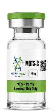
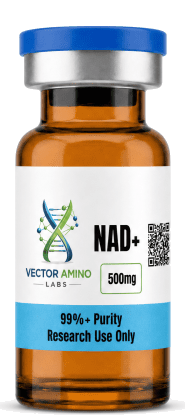

In the rapidly advancing field of metabolic and longevity research, few discoveries have generated as much excitement as Mitochondrial-Derived Peptides (MDPs). Among these, MOTS-c (Mitochondrial Open Reading Frame of the 12S rRNA-c) has emerged as a profound compound of interest, frequently referred to in research literature as an “exercise mimetic.”
For biomedical researchers investigating cellular bioenergetics, insulin resistance, and age-related metabolic decline, MOTS-c represents a fundamental shift in our understanding of mitochondrial function. This comprehensive guide explores the molecular mechanisms, AMPK activation pathways, and latest quantitative research surrounding MOTS-c in controlled experimental settings.
Disclaimer: The compounds discussed in this article are intended strictly for laboratory research and development purposes. They are not approved for human or animal consumption, nor are they intended to address any disease. All products are intended for laboratory and educational use by qualified professionals only.
The Discovery of Mitochondrial-Derived Peptides
Historically, mitochondria were viewed simply as the “powerhouses of the cell,” organelles responsible solely for generating adenosine triphosphate (ATP) through oxidative phosphorylation. However, recent research has revealed that mitochondria also function as critical signaling organelles, actively communicating with the cell nucleus to regulate metabolism and cellular homeostasis.
This communication is facilitated, in part, by Mitochondrial-Derived Peptides. MOTS-c is a unique 16-amino-acid peptide that is encoded not within the cell’s nuclear DNA, but within the mitochondrial genome itself [1]. Upon translation, MOTS-c translocates from the mitochondria to the nucleus, where it actively regulates the expression of genes involved in metabolic homeostasis and stress response.
Mechanism of Action: The AMPK Pathway
The primary mechanism of action for MOTS-c centers on its profound ability to activate AMP-activated protein kinase (AMPK), often described as the master metabolic switch of the cell [2].
In laboratory models, MOTS-c research application has been shown to significantly increase the levels of 5-aminoimidazole-4-carboxamide-1-β-D-ribofuranoside (AICAR), a known activator of AMPK. The activation of the AMPK pathway triggers a cascade of metabolic adaptations designed to restore cellular energy balance:
- Enhanced Glucose Uptake: MOTS-c stimulates the translocation of GLUT4 transporters to the cell membrane, significantly increasing glucose uptake in skeletal muscle independent of insulin signaling [3].
- Increased Fatty Acid Oxidation: By activating AMPK, MOTS-c promotes the breakdown of fatty acids for energy, reducing lipid accumulation in the liver and adipose tissue.
- Mitochondrial Biogenesis: MOTS-c upregulates the expression of PGC-1α, driving the formation of new, healthy mitochondria and improving overall cellular respiratory capacity.
The “Exercise Mimetic” Effect in Preclinical Models
Because physical exercise naturally activates the AMPK pathway, compounds that can artificially stimulate this pathway are heavily researched as potential “exercise mimetics.” In preclinical animal models, MOTS-c has demonstrated remarkable efficacy in replicating many of the metabolic benefits of physical training.
Recent 2025 and 2026 studies have highlighted the profound impact of MOTS-c on insulin resistance and obesity. In diet-induced obesity models, mice treated with MOTS-c exhibited significantly reduced weight gain and improved glucose tolerance compared to control groups, despite consuming the same high-fat diet [4]. Furthermore, research indicates that MOTS-c research application can enhance physical performance and exercise capacity in aging animal models, suggesting a potential role in combating age-related metabolic decline and sarcopenia [5].
Comparison: MOTS-c vs. NAD+
When designing metabolic research protocols, investigators frequently compare or combine MOTS-c with NAD+ precursors. The following table outlines the distinct mechanisms of these two prominent metabolic compounds.
| Compound | Primary Source/Origin | Primary Mechanism of Action | Key Metabolic Effect |
|---|---|---|---|
| MOTS-c | Mitochondrial DNA (12S rRNA) | Direct activation of AMPK pathway | Acts as an exercise mimetic, enhancing glucose uptake and fatty acid oxidation |
| NAD+ | Cellular Coenzyme | Essential co-factor for sirtuins and PARPs | Facilitates electron transport and cellular DNA repair |
It is important to note that these compounds are highly synergistic. Research indicates that MOTS-c increases cellular NAD+ levels as part of its downstream mechanism, making the study of their combined effects a highly active area of investigation [6].
Conclusion for Laboratory Professionals
MOTS-c represents a critical frontier in the study of cellular bioenergetics and metabolic regulation. By acting as an intracellular signaling molecule that bridges the mitochondria and the nucleus, it provides researchers with a powerful tool for investigating insulin resistance, obesity, and age-related metabolic dysfunction.
For laboratories requiring premium, third-party tested compounds, Vector Amino Labs provides research-grade peptides with verified Certificates of Analysis (COA) to ensure absolute precision and reliability in your experimental protocols.
References
[1] “MOTS-c: A promising mitochondrial-derived peptide for therapeutic applications.” PubMed Central.[2] “Mitochondrial-Encoded Peptide MOTS-c, Diabetes, and Aging.” Diabetes & Metabolism Journal, 2023.
[3] “Mitochondria-derived peptide MOTS-c: effects and mechanisms.” PubMed Central, 2023.
[4] “MOTS-c Peptide: 44% Drop in Insulin Resistance.” Metabolic Research Institute, 2026.
[5] “Effect of aerobic and resistance exercise on the mitochondrial peptide MOTS-c.” Nature Scientific Reports, 2021.
[6] “MOTS-c vs NAD+: Benefits and Key Differences.” Perfect B, 2026.
This content is provided for educational and informational purposes only, summarizing published peer-reviewed research. All compounds referenced are intended exclusively for in-vitro laboratory research and are not intended, labeled, or approved for human use.